3D breast imaging technique improves cancer detection rate
(Via Medical News Today) A study published in JAMA has found that the addition of tomosynthesis, a 3D imaging technique, to digital mammography was linked to a reduction in the number of patients being called back for additional testing and a rise in the breast cancer detection rate.
Digital tomosynthesis is a relatively new imaging technique that was only approved by the US Food and Drug Administration (FDA) in 2011 and has yet to become widely available.
Mammograms are currently the main method of screening forbreast cancer. Breastcancer.org reports that mammograms have been shown to lower the risk of dying from breast cancer by 35% in women over the age of 50. Last year, approximately 39,620 women were expected to die from breast cancer in the US.
But the standard digital mammogram has been observed to have its limitations. It has been criticized for producing too many false-positive results, having limited sensitivity and for, at times, over-diagnosing clinically insignificant lesions.
The compression of the breast can also be uncomfortable and occasionally hide lesions in overlapping tissue that will not appear on a mammogram.
A new method of screening
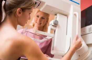
Digital tomosynthesis aims to address these problems by taking multiple X-ray pictures from different angles. The breast is positioned as it is for a conventional mammogram, but only a small amount of pressure is applied. The information is used to produce 3-D images throughout the entire breast, instead of a single one that conventional mammograms do.
Dr. Sarah M. Friedewald, of Advocate Lutheran General Hospital, Park Ridge, IL, and her team set out to determine whether mammography combined with tomosynthesis is associated with better performance of American breast screening programs.

Tomosynthesis is a new imaging technique that doctors hope can overcome the limitations of conventional mammograms.
Using data from 13 centers, the team evaluated a total of 454,850 examinations, of which 173,663 utilized a combination of digital mammography and tomosynthesis.
The study recorded the number of patients recalled for further imaging, cancer detection rate, the number of patients recalled following a diagnosis of breast cancer and the number of patients who underwent biopsies following a diagnosis of breast cancer.
The utilization of digital tomosynthesis resulted in an associated reduction in the recall rate, from 107 to 91 per 1,000 screens. Cancer detection rose from 4.2 to 5.4, and for invasive cancer specifically this rise was from 2.9 to 4.1 cases per 1,000 screens.
The addition of tomosynthesis also increased the positive predictive value – the number of patients found to have the disease – for recall from 4.3% to 6.4%, and for biopsy it increased from 24.2% to 29.2%.
The authors conclude that the addition of tomosynthesis to digital mammography was associated with a decrease in the rate of recall and an increase in the cancer detection rate. They say:
“The association with fewer unnecessary tests and biopsies, with a simultaneous increase in cancer detection rates, would support the potential benefits of tomosynthesis as a tool for screening. However, assessment for a benefit in clinical outcomes is needed.”
Dr. Friedewald believes that the study provides a compelling story about the effectiveness of 3D mammography as it is 10 times larger than previous studies and because the data came from both academic and community health care settings.
The ‘screening debate’
An editorial piece in JAMA linked to the study agrees on the likelihood of tomosynthesis being an advancement over mammography, saying that “tomosynthesis potentially improves sensitivity while addressing the two most common arguments against mammography screening – false-positive findings and over diagnosis.”
However, they say that fundamental questions about screening remain. With regard to this study in particular, the editors write that the research is hampered by a lack of long-term follow-up information and the fact that new technology has made the 2-view digital mammography used in the study obsolete.
Reference is also made to eight randomized clinical trials of screening mammography that failed to find a reduction in cancer-related mortality as a result the screening process, spurring on debate over the benefits of breast cancer screening.
In their editorial, Dr. Etta Pisano, of the Medical University of South Carolina in Charleston, and Martin Yaffe, Ph.D., of the University of Toronto, call for the National Institutes of Health (NIH) to fund a multi-site clinical trial of modern technology to provide an end to the screening debate once and for all:
“The continuing controversy surrounding the most effective strategy for deploying the various available technologies continues unabated, and clear consensus is lacking on when to screen, how often, and with what tools, or even which screen-detected cancers could be managed more conservatively.”
Recently, Medical News Today reported on a study that found there were limitations in the conventional way in which doctors classify breast cancer tumors, leading to incorrect classification and patients missing out on treatment.
Written by James McIntosh


